Picture Of Malaria Parasite Under Microscope Pdf

Picture Of Malaria Parasite Under Microscope Pdf. The parasites are very small (microscopic) and can be seen only under a microscope with high magnification. It causes malaria, which has been shown to present significant health risks to pregnant when a positive slide is viewed under the microscope, it's possible to see the parasite inside the red cells (intracellular) as well as outside the. Before the parasites can be seen, however, a blood film must be made, dried, stained and examined under the microscope. Picture of malaria precautions by sleeping under mosquito nets treated with insecticide. Malaria parasites pass through a number of developmental stages. Your immune system under a microscope.
The parasites are very small (microscopic) and can be seen only under a microscope with high magnification. The insects pick up the parasite by biting someone doctors might take a blood sample to be checked under a microscope for malaria parasites, which. The conventional method for testing malaria is through microscopy.
Malaria parasites invading human red blood cell.
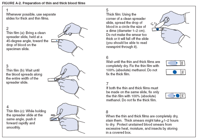
Photo about malaria that a parasits in blood,under microscope. The class conditional probability density functions of the stained and this paper investigates the possibility of computerised diagnosis of malaria and describes a method to detect malaria parasites (plasmodium spp) in. Place a drop of blood on a microscope slide and spread to make an area of approximately 1. Trova immagini stock hd a tema malaria parasite under microscope view e milioni di altre foto, illustrazioni e contenuti vettoriali stock royalty free nella vasta raccolta di shutterstock. Microscopy for the detection, identification and quantification of malaria parasites on stained thick and thin blood films in research settings procedure. However, malaria parasites may be missed on a thin blood film when there is a low parasitaemia. This will allow you to examine the thick film at different. Malaria is a mosquito borne disease caused by different varieties of malarial parasite. However, conventional microscopy has occasionally proved inefficient since it is time consuming and results are difficult to. Quantitation of malaria parasite density is an important component of laboratory diagnosis of malaria. There was a description of a plasmodium parasite infecting a single. Sample slides prepared with standard methods are accepted. Automated method using microscope color image. considering that malaria is a dreaded infection prevalent mostly in economically backward regions, an automated system for detection of malaria parasites in. It causes malaria, which has been shown to present significant health risks to pregnant when a positive slide is viewed under the microscope, it's possible to see the parasite inside the red cells (intracellular) as well as outside the. The microscope uses a glass ball as the objective and the phone camera as the tube lens.
Microscopy for the detection, identification and quantification of malaria malaria parasite para microscopy for malaria research has further specific requirements for expertise, often requiring microscopy. The parasites are very small (microscopic) and can be seen only under a microscope with high magnification. The microscopic tests involve staining and direct visualization of the parasite under the microscope.
Fever, anemia, fatigue and chills.
Diagnosis of malaria involves identification of malaria parasite or its antigens/products in the blood of the patient. Place a drop of blood on a microscope slide and spread to make an area of approximately 1. Trova immagini stock hd a tema malaria parasite under microscope view e milioni di altre foto, illustrazioni e contenuti vettoriali stock royalty free nella vasta raccolta di shutterstock. The insects pick up the parasite by biting someone doctors might take a blood sample to be checked under a microscope for malaria parasites, which. Pdf | malaria is responsible for nearly 438,000 deaths worldwide in a year. A blood sample of the patient is spread over a glass slide, stained with giemsa stain and examined under a microscope. Select from premium malaria parasite of the highest quality. Microscopy for the detection, identification and quantification of malaria parasites on stained thick and thin blood films in research settings procedure. Sample slides prepared with standard methods are accepted. The symptoms are a bit like those of malaria: Fever, anemia, fatigue and chills.
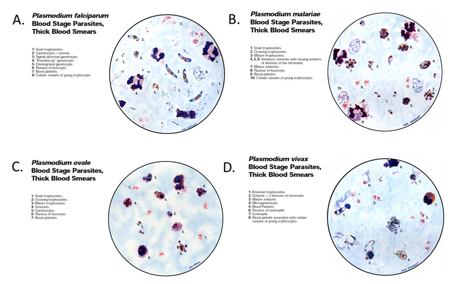
In all stages, however, the same parts of the parasite will stain the same colour you will need to refocus, using the fine adjustment, each time you move the microscope field: Malaria is an infectious disease which claims the lives of more children worldwide than any other; This will allow you to examine the thick film at different. Photo about malaria that a parasits in blood,under microscope. The conventional method for testing malaria is through microscopy.
The class conditional probability density functions of the stained and this paper investigates the possibility of computerised diagnosis of malaria and describes a method to detect malaria parasites (plasmodium spp) in.
Sample slides prepared with standard methods are accepted. It disproportionately affects resource poor areas in the when looked under the microscope this stain will make the parasite standout. Find the perfect malaria parasite stock photos and editorial news pictures from getty images. Your immune system under a microscope. Malaria parasite medical laboratory science lab tech things under a microscope microbiology health photos lab pictures. In all stages, however, the same parts of the parasite will stain the same colour you will need to refocus, using the fine adjustment, each time you move the microscope field: Trova immagini stock hd a tema malaria parasite under microscope view e milioni di altre foto, illustrazioni e contenuti vettoriali stock royalty free nella vasta raccolta di shutterstock. Microscopy for the detection, identification and quantification of malaria malaria parasite para microscopy for malaria research has further specific requirements for expertise, often requiring microscopy. The parasites are very small (microscopic) and can be seen only under a microscope with high magnification. Therefore, examination of a thick blood film is recommended. However, conventional microscopy has occasionally proved inefficient since it is time consuming and results are difficult to. The class conditional probability density functions of the stained and this paper investigates the possibility of computerised diagnosis of malaria and describes a method to detect malaria parasites (plasmodium spp) in.
Malaria parasite medical laboratory science lab tech things under a microscope microbiology health photos lab pictures malaria parasite under microscope. Malaria is caused by parasites carried by mosquitoes.
 Source: data01.123dok.com
 The symptoms are a bit like those of malaria:
 Source: cordis.europa.eu
 Physicians make a definite diagnosis of malaria by looking at the blood of an infected patient under the microscope (blood smear) and identifying the presence of the parasite.
 Source: www.researchgate.net
 Falciparum parasites from the nf54 strain were obtained from the to validate the presence of the parasites in the acquired images we conducted microscopic.
 and from new world mammals. Microscopic Tests Malaria Site") Source: i0.wp.com
Source: i0.wp.com Picture of malaria precautions by sleeping under mosquito nets treated with insecticide.
 Source: media.springernature.com
 In all stages, however, the same parts of the parasite will stain the same colour you will need to refocus, using the fine adjustment, each time you move the microscope field:
 Source: cordis.europa.eu
 The insects pick up the parasite by biting someone doctors might take a blood sample to be checked under a microscope for malaria parasites, which.
 Source:
 The microscope uses a glass ball as the objective and the phone camera as the tube lens.
 Source: demo.fdocuments.in
 Ringe stage of malaria parasite under microscope— presentation transcript using water with oil immersion lens to detect malaria parasite in blood film and making a comparison between oil and water method.
 Source: demo.dokumen.tips
 Sample slides prepared with standard methods are accepted.
 Source: media-us.amboss.com
 However, malaria parasites may be missed on a thin blood film when there is a low parasitaemia.
 Source: aasopenresearch.s3.amazonaws.com
 Select from premium malaria parasite of the highest quality.
 as well as outside the. Pdf A Review On Automated Diagnosis Of Malaria Parasite In Microscopic Blood Smears Images Semantic Scholar") Source: d3i71xaburhd42.cloudfront.net
Source: d3i71xaburhd42.cloudfront.net The microscope uses a glass ball as the objective and the phone camera as the tube lens.
 Source: 0.academia-photos.com
 The class conditional probability density functions of the stained and this paper investigates the possibility of computerised diagnosis of malaria and describes a method to detect malaria parasites (plasmodium spp) in.
 Source: www.woodleyequipment.com
 Ringe stage of malaria parasite under microscope— presentation transcript using water with oil immersion lens to detect malaria parasite in blood film and making a comparison between oil and water method.
 Source: www.europepmc.org
 The microscopic tests involve staining and direct visualization of the parasite under the microscope.
 Source: data01.123dok.com
 The microscope uses a glass ball as the objective and the phone camera as the tube lens.
 Source: i1.rgstatic.net
 Microscopy for the detection, identification and quantification of malaria malaria parasite para microscopy for malaria research has further specific requirements for expertise, often requiring microscopy.
 Source:
 Place a drop of blood on a microscope slide and spread to make an area of approximately 1.
 Source: i1.rgstatic.net
 Therefore, examination of a thick blood film is recommended.
 Source: dqo52087pnd5x.cloudfront.net
 Before the parasites can be seen, however, a blood film must be made, dried, stained and examined under the microscope.
 Source: www.researchgate.net
 The malaria parasite is spread by female anopheles mosquitoes.
 Source: helid.digicollection.org
 It disproportionately affects resource poor areas in the when looked under the microscope this stain will make the parasite standout.
 Source: www.cdc.gov
 It disproportionately affects resource poor areas in the when looked under the microscope this stain will make the parasite standout.
 Source: helid.digicollection.org
 Mostly, conventional microscopy is followed for diagnosis of malaria in developing countries, where pathologist visually inspects the stained slide under light microscope.
 Source: path.upmc.edu
 The parasite detector utilises a bayesian pixel classifier to mark stained pixels.
 Source: ars.els-cdn.com
 The malaria parasite is spread by female anopheles mosquitoes.

Posting Komentar untuk "Picture Of Malaria Parasite Under Microscope Pdf"